Table of Contents
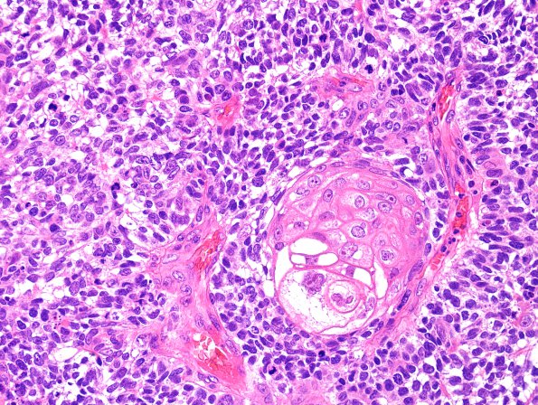
Washington University Experience | NEOPLASMS (METASTASES) | Microscopic | 75A2 Metastasis, lung, squamous (Case 75) H&E 2.jpg
Sections of the left frontal lobe mass show sheets of cohesive tumor cells with multiple areas of keratinization and other areas that appear almost basaloid with nuclear palisading.

