Table of Contents
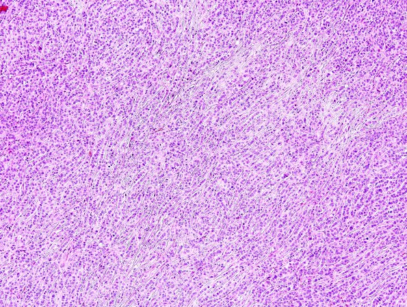
Washington University Experience | NEOPLASMS (METASTASES) | Microscopic | 77A1 Metastasis, breast (Case 77) H&E 2.jpg
77A1,2 The neoplastic cells are arranged in variable patterns (single-file, solid nests and glandular/ductular) with scattered dystrophic calcifications. The individual tumor cells are characterized by scant to moderate amount of eosinophilic cytoplasm, nuclei that vary from round to ovoid to significantly atypical, showing enlargement, irregular nuclear membranes, and scattered examples of multinucleation. The chromatin patterns vary from finely dispersed with small chromocenters, to cells with prominent nucleoli to hyperchromatic forms. Mitotic figures are readily identified. Additional findings include focal invasion of adjacent brain parenchyma, patchy chronic inflammatory cell infiltrate, multiple vessels with fibrinoid necrosis and few fragments of uninvolved cerebral cortex and white matter.

