Table of Contents
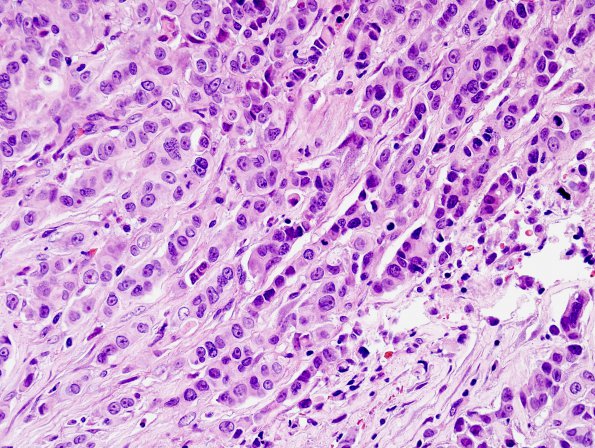
Washington University Experience | NEOPLASMS (METASTASES) | Microscopic | 77A2 Metastasis, breast (Case 77) H&E 1.jpg
The neoplastic cells are arranged in variable patterns (single-file, solid nests and glandular/ductular) with scattered dystrophic calcifications. ---- Not shown: Immunohistochemical stains show the majority of her tumor cells to be positive for mammaglobin and estrogen receptor is expressed by majority of tumor cell nuclei with intensity ranging from weak to strong. Progesterone receptor is negative. A HER-2 immunostain is equivocal with 2+ reactivity; We also reviewed representative sections from her prior needle biopsy and the tumors are histologically comparable. The overall findings are consistent with a metastatic carcinoma from her known breast primary.

