Table of Contents
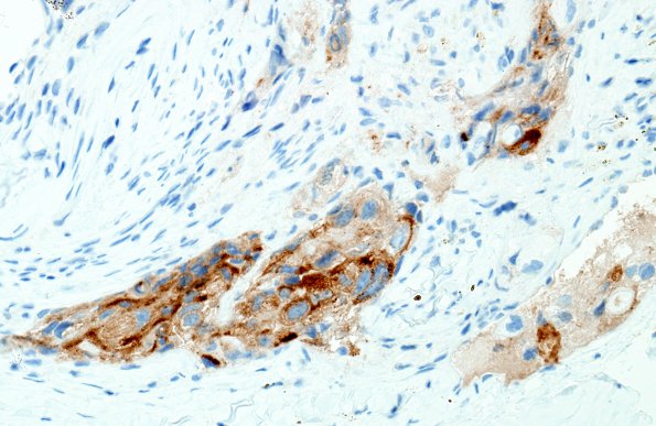
Washington University Experience | NEOPLASMS (METASTASES) | Microscopic | 7B Metastasis, prostate (Case 7) PSA 1
Immunohistochemistry was performed to ascertain the site of origin of the metastasis. The tumor cells are positive for prostate specific antigen (PSA, shown) and prostatic acid phosphatase (PSAP). The tumor nests also demonstrate weak AMACR and renal cell carcinoma antigen (RCC) staining, however, both of these are nonspecific and can be seen in adenocarcinoma of prostatic origin. There is also focal CK20 positivity, which has been described in prostatic adenocarcinoma. CK7 and thrombomodulin are negative. The tumor cells also demonstrate diffuse CD10 positivity. Overall, the tumor morphology and strong PSA and PSAP staining are consistent with a metastasis from the patient's known prostate cancer.

