Table of Contents
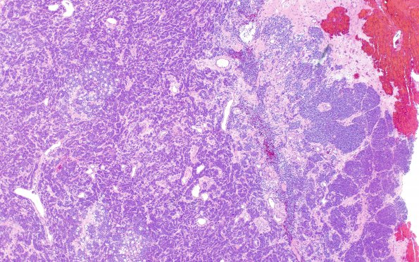
Washington University Experience | NEOPLASMS (METASTASES) | Microscopic | 87A1 Metastasis, breast (Case 87) H&E 4
Case 87 History ---- The patient is a 73-year-old woman with invasive lobular carcinoma diagnosed in 2012 and right axilla lymph node metastasis, who has a 2.8 x 1.8 cm left frontal dura-based lesion and a nearby separate punctate lesion. Operative procedure: Craniotomy left frontal for tumor resection. ---- 87A1-3 Sections show a metastatic carcinoma with extensive necrosis. The tumor cells have vesicular chromatin, one to multiple prominent nucleoli, and moderate amounts of eosinophilic cytoplasm. In some areas, the tumor cells appear discohesive, and are invading as files of single cells. In other areas, the tumor cells are arranged in cords or small nests. There are numerous mitotic figures and karyorrhectic debris. The tumor cells in the current specimen seemed to show higher nuclear grade than the patient's prior biopsy specimen.

