Table of Contents
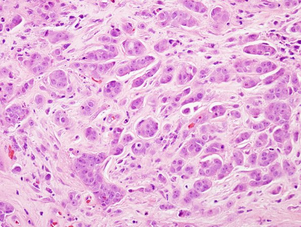
Washington University Experience | NEOPLASMS (METASTASES) | Microscopic | 88A2 Metastasis, breast (Case 88) H&E 1
H&E shows an infiltrative tumor that is composed of sheets, nests, single file infiltrative growth and poorly formed glandular/ductal structures. The tumor-brain interface is relatively well defined, favoring a metastasis. ---- Not Shown: A panel of immunohistochemical stains was performed and included ER, PR, Her2/Neu, CK7, CK20 and mammaglobin. Mammaglobin highlighted select stromal areas of infiltrative tumor growth. Loss of breast markers is not uncommon in the metastatic setting. Based on the patient's known prior history of breast cancer, the histologic and immunohistologic features, a diagnosis of metastatic carcinoma is most consistent with a breast primary.

