Table of Contents
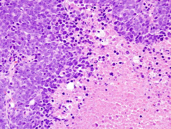
Washington University Experience | NEOPLASMS (METASTASES) | Microscopic | 8A Metastasis, prostate (Case 8) H&E 4
Case 8 History ---- The patient is a 76-year-old man with a history of prostate cancer who presents with lower extremity weakness. CT of the thoracic spine shows a soft tissue mass in the right aspect of the spinal canal at T3-T4, a soft tissue lesion in the left L5 pedicle, and a sclerotic lesion at the superior endplate of L4. Operative procedure: Posterior spinal fusion with resection of epidural mass. ---- 8A Sections show malignant epithelioid cells growing in sheet-like fashion that form large nests separated by bands of eosinophilic collagenous tissue. The large nests have central necrotic cores filled with neoplastic 'ghost'-cells. The neoplastic cells have large round to ovoid shaped nuclei with smudged chromatin and prominent eosinophilic nucleoli. They have small to moderate amounts of eosinophilic cytoplasm. Mitotic figures are easily identified and number up to 8 mitoses in a single high power field.

