Table of Contents
Washington University Experience | NEOPLASMS (METASTASES) | Microscopic | 91A Metastasis, breast (Case 91) HER-2 5.jpg
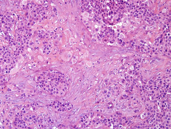
Case 91 History ---- The patient is a 59-year-old woman with a history of breast carcinoma in 2011, presenting one year later with two enhancing brain masses. Operative procedure: Left parietal craniotomy for tumor excision. ---- 91A This mass is comprised of epithelioid tumor cells arranged in sheets, nests and single scattered large, atypical cells with abundant tumoral necrosis and desmoplastic reaction. Most of the tumor cells are relatively uniform with abundant eosinophilic cytoplasm, clear cell border, round to oval nuclei with finely stippled chromatin and prominent central nucleoli. Singly scattered bizarre cells are also present. Mitotic figures are frequent, including atypical forms.

