Table of Contents
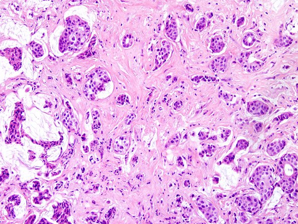
Washington University Experience | NEOPLASMS (METASTASES) | Microscopic | 93A1 Metastasis, breast & RadioRx pituitary (Case 93) H&E 1.jpg
93A1-4 Sections of the "right frontal tumor" and "suprasellar tumor" both show a neoplastic proliferation of epithelioid cells growing in a solid pattern with predominantly nested architecture, with a few, rare, scattered tubules. The surrounding cortical tissues are reactive, edematous, and partially necrotic. Many of the blood vessels show prominent eosinophilic hyalinization, consistent with the patient's history of whole brain radiation in 2012. The tumor cells themselves are characterized by moderate amounts of eosinophilic cytoplasm, enlarged, ovoid nuclei with prominent nucleoli, mild to moderate nuclear contour irregularities, and a generally finely granular pattern of chromatin distribution. The nuclear to cytoplasmic ratio is increased. At low magnification, the cells appear somewhat monomorphic. Mitotic activity is markedly increased, with > 30 mitotic figures/10 HPF.

