Table of Contents
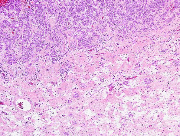
Washington University Experience | NEOPLASMS (METASTASES) | Microscopic | 93A2 Metastasis, breast & RadioRx (Case 93) H&E 5.jpg
Sections of the "right frontal tumor" and "suprasellar tumor" both show a neoplastic proliferation of epithelioid cells growing in a solid pattern with predominantly nested architecture, with a few, rare, scattered tubules. The surrounding cortical tissues are reactive, edematous, and partially necrotic. Many of the blood vessels show prominent eosinophilic hyalinization, consistent with the patient's history of whole brain radiation in 2012.

