Table of Contents
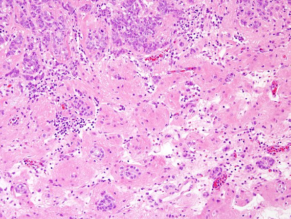
Washington University Experience | NEOPLASMS (METASTASES) | Microscopic | 93A3 Metastasis, breast & RadioRx (Case 93) H&E 4.jpg
Sections of the "right frontal tumor" and "suprasellar tumor" both show a neoplastic proliferation of epithelioid cells growing in a solid pattern with predominantly nested architecture, with a few, rare, scattered tubules. The surrounding cortical tissues are reactive, edematous, and partially necrotic. (H&E)

