Table of Contents
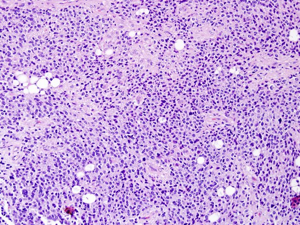
Washington University Experience | NEOPLASMS (NEURONAL) | Central Neurocytoma | 10A5 Liponeurocytoma, cerebellar (Case 10) H&E 10.jpg
10A5-8 In some areas, there are large clear vacuoles reminiscent of fat cells or lipid accumulation. Only occasional mitotic figures are evident. However, there is focal microvascular proliferation. In some areas, the tumor cells are arranged around delicate fibrillary material, consistent with neuropil. These structures resemble neurocytic rosettes.

