Table of Contents
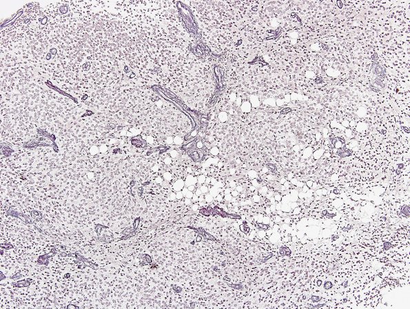
Washington University Experience | NEOPLASMS (NEURONAL) | Central Neurocytoma | 10F Liponeurocytoma, cerebellar (Case 10) Retic 1.jpg
A histochemical stain for reticulin highlights intratumoral vessels, however, there is minimal deposition around tumor cells. ---- Ancillary studies (not shown) ---- A stain for neurofilament protein highlights entrapped axons at the periphery of the neoplasm. However, tumor cells are negative. The tumor is also negative for epithelial membrane antigen. Given the inclusion of oligodendroglioma in the differential diagnosis, genetic studies were pursued. FISH showed polysomies (gains) of both chromosomes 1 and 19. No deletions were found. This genetic pattern likely reflects an overall state of aneuploidy/polyploidy and is non-specific. ---- A diagnosis of Cerebellar Liponeurocytoma, WHO Grade 2 was made. This is a rare tumor and I have included it with the central neurocytomas which it resembles. The WHO CNS 2021 Bluebook separates this tumor from typical central neurocytomas but with molecular profiles more similar to those of central neurocytoma than medulloblastoma, Nonetheless, cerebellar liponeurocytoma displays a unique epigenetic signature. The lipid-laden cells represent lipid accumulation in neuroepithelial tumor cells. Focal GFAP expression by tumor cells are commonly observed. This tumor has a favorable prognosis.

