Table of Contents
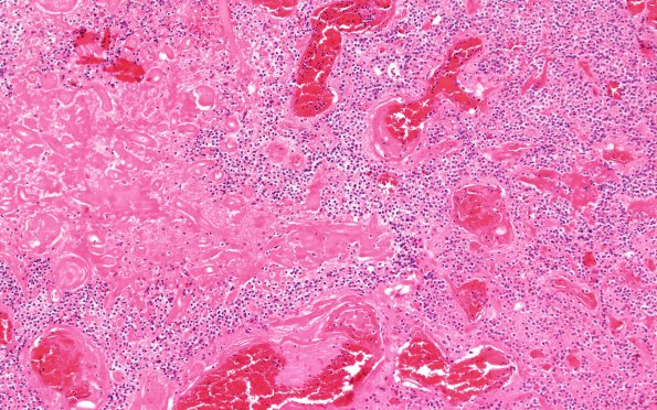
Washington University Experience | NEOPLASMS (NEURONAL) | Central Neurocytoma | 11A2 Central Neurocytoma (Case 11) 10X
Occasionally there are areas of necrosis and hyalinized vasculature (H&E) ---- Ancillary studies (not shown): Immunohistochemical stains performed show that the cells are diffusely positive for synaptophysin. GFAP shows a high background positivity which made the stain non-interpretable. Scattered cells showed faint nuclear positivity for NeuN. A diagnosis of central neurocytoma was made.

