Table of Contents
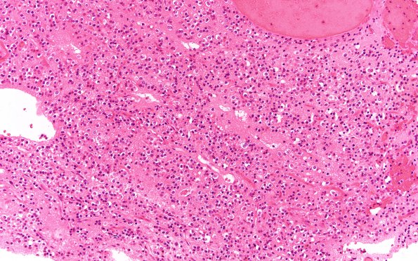
Washington University Experience | NEOPLASMS (NEURONAL) | Central Neurocytoma | 12A Neurocytoma, central (Case 12) H&E 20X
The patient is a 43 year old man who presented in April of 2004 with headaches and was found to have a large intraventricular mass diagnosed as a central neurocytoma. He underwent a transcallosal resection and shunt placement. The mass recurred and in May of 2005 he underwent re-resection and shunt revision. Since this time the patient has received fractionated radiation therapy, stereotactic radiosurgery, and, in August 2010, presented with a recurrent lesion. ---- The 2005 neurosurgical specimen is shown in the following images: Sections show a hypocellular to moderately cellular neoplasm whose neoplastic cells have minimal cytoplasm with clear pericellular halos around them. The nuclei are round with smooth nuclear membranes for most parts. In some areas, the tumor cells are embedded in a background of neuropil. Mitoses are rare. Microvascular proliferation is not seen.

