Table of Contents
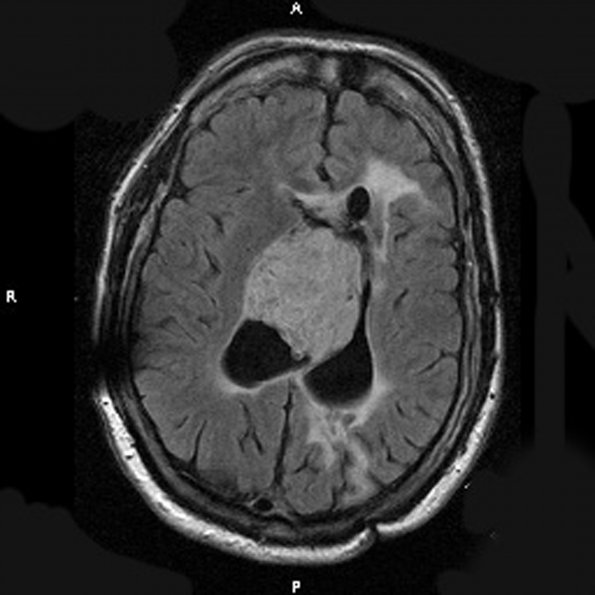
Washington University Experience | NEOPLASMS (NEURONAL) | Central Neurocytoma | 3A1 Neurocytoma, central (Case 3) T1W A - Copy
Case 3 History ---- The patient was a 60 year old man with a history of an intraventricular tumor who initially presented at the age of 35 to an outside hospital in 1986 with memory issues and confusion. He underwent resection and his tumor was initially diagnosed as an ependymoma. He was subsequently found by CT to have a large exophytic intraventricular tumor occupying most of both frontal horns. It showed extension down to and probably into the foramina of Monro and possibly into the anterior third ventricle. It also showed amorphous clumps of calcification. In 2001 the patient had recurrent tumor re-resected. The material corresponding to this resection was reviewed at BJH in-house and diagnosed as a central neurocytoma, WHO grade II; it was noted that the lesion had many low-power features mimicking an ependymoma. Operative procedure: Left frontal craniotomy with resection of tumor. ---- 3A1-3 MRI Images represent scans before the 2011 resections which show a mixed cystic and solid mass centered within the septum pellucidum. ---- 3A1 This scan is a T1-weighted scan with contrast administered.

