Table of Contents
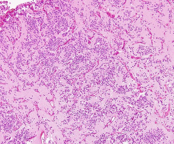
Washington University Experience | NEOPLASMS (NEURONAL) | Central Neurocytoma | 3B1 Neurocytoma, central (Case 3) H&E 1
3B1-5 Sections show a hypercellular glial neoplasm with a low magnification appearance consisting of sheets of neoplastic cells with a nodular growth pattern that are separated by areas of acellular neuropil. Low magnification examination also showed the presence of numerous small vessels that are often separated from neoplastic cells by a rim of acellular neuropil. Neoplastic cells have round nuclei with smooth nuclear envelopes and mildly stippled hyperchromatic chromatin. Many of the cells have thin rims of mildly eccentric eosinophilic cytoplasm and others have indistinct cell bodies. Perivascular neuropil is unoriented and structureless in some areas but, in others forms perivascular pseudorosettes but no 'true' ependymal rosettes. Mitotic figures are not observed.

