Table of Contents
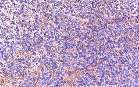
Washington University Experience | NEOPLASMS (NEURONAL) | Central Neurocytoma | 8B Neurocytoma, central (Case 8) SYN 40X
Immunoperoxidase stains show that the cells are positive for synaptophysin. ---- Comment: “The main differential considered on this case based on the H&E appearance of the tumor was oligodendroglioma and central neurocytoma, which may be difficult to distinguish with certainty on H&E alone. However, characteristics of this tumor arguing for the diagnosis of central neurocytoma include the high cellularity with very little pleomorphism. Finally, the tumor appears to be well demarcated from the surrounding brain parenchyma, in contrast to the usual infiltrative pattern demonstrated by oligodendroglial tumors. The pattern of immunoperoxidase staining also supports the diagnosis of central neurocytoma. The tumor cells are with only very few exceptions S100 negative, although oligodendroglial cells in the adjacent normal parenchyma stained strongly with S100. Though NSE is somewhat nonspecific, the uniformly intense staining for this marker exhibited by the tumor argues for neuronal differentiation. Given that the clinical setting appears to be appropriate for central neurocytoma, we believe that this is the most appropriate diagnosis.”

