Table of Contents
Washington University Experience | NEOPLASMS (NEURONAL) | Cerebellar Liponeurocytoma | 1C2 Liponeurocytoma, cerebellar (Case 1) NF 4.jpg
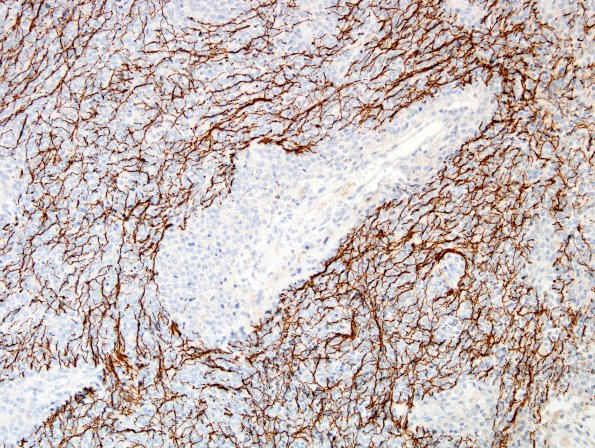
A stain for neurofilament protein highlights areas of diffuse infiltration with other regions showing a solid pattern of growth (NF IHC). However, tumor cells themselves are negative.

