Table of Contents
Washington University Experience | NEOPLASMS (NEURONAL) | Cerebellar Liponeurocytoma | 1E1 Liponeurocytoma, cerebellar (Case 1) GFAP 8.jpg
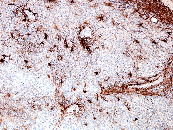
1E1,2 A stain for GFAP highlights numerous elongate cytoplasmic processes, likely entrapped elements; however, scattered tumor cells may be GFAP immunoreactive. (GFAP IHC)

