Table of Contents
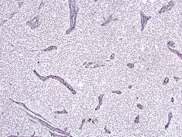
Washington University Experience | NEOPLASMS (NEURONAL) | Cerebellar Liponeurocytoma | 1G Liponeurocytoma, cerebellar (Case 1) Retic 4.jpg
A reticulin stain only highlights the vascularity. (Reticulin histochemistry) ---- Not shown: The tumor is also negative for epithelial membrane antigen. Strong nuclear staining for Neu-N is seen within a subset of tumor cells. Given the inclusion of oligodendroglioma in the differential diagnosis, FISH studies were performed and showed polysomies (gains) of both chromosomes 1 and 19. No deletions were found. ---- Comment: The morphologic, immunohistochemical, and genetic features are consistent with the diagnosis of cerebellar liponeurocytoma, WHO grade 2. Although the clinical behavior of such cases is generally favorable, recurrences are common.

