Table of Contents
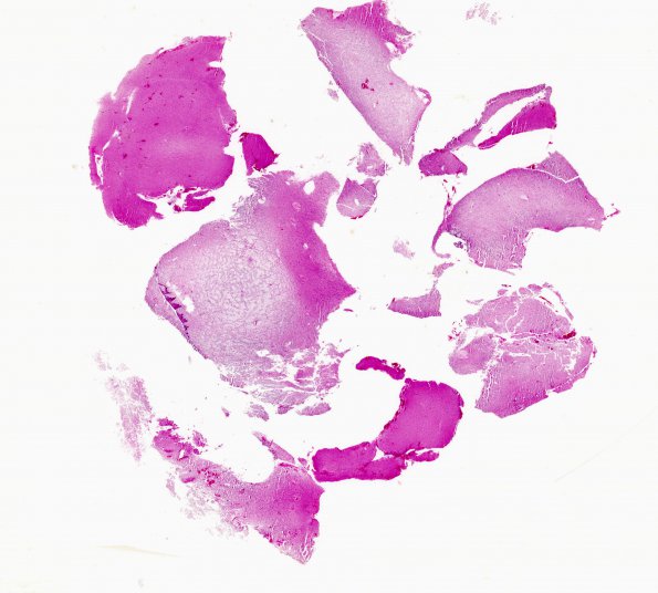
Washington University Experience | NEOPLASMS (NEURONAL) | DNET | 10A1 DNET (Case 10) H&E whole mount
Case 10 History ---- The patient is a 16 year old boy who experienced a single episode of seizures. Imaging shows a non-enhancing mass adjacent to the sensory cortex. Clinical diagnosis: Low grade glioma. Operative procedure: Craniotomy: excision of parietal mass. ---- 10A1-4-Sections of the right parietal lesion show a glioneuronal neoplasm consisting of columns of oligodendroglia-like cells oriented perpendicular to the cortical surface in a pale mucinous to eosinophilic matrix with microcystic areas. There are scattered "floating" neurons in these spaces. There are scattered neurons in the adjacent white matter. Mitoses are hard to find.

