Table of Contents
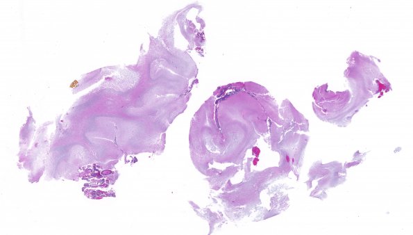
Washington University Experience | NEOPLASMS (NEURONAL) | DNET | 13A1 DNET, cerebellum (Case 13) H&E whole mount
Case 13 History ---- The patient is a 46 year old man with a previous history of diplopia and progressive hearing loss in 2007. The patient underwent excisional biopsy of the left cerebellum in September of 2007 and was diagnosed with a DNT. Per clinical records, he has received no adjuvant therapy and during a visit on 7/16/2010 there was a question of an evolving focus of enhancement at the pontomedullary junction. ---- 13A1-4 The cerebellum was involved by a low grade glioneuronal neoplasm. The neoplasm forms nodular areas containing strands of cells organized with an architecture of columns that appear to float within a blue myxoid background. The neoplastic cells have small nuclei with hyperchromatic chromatin and many show peri-nuclear cytoplasmic clearing. Occasional neurons can also be seen 'floating' within the myxoid background material. The involvement appears patchy with foci of neoplasm separated by more normal appearing cerebellum. Mitotic figures are not appreciated. Endothelial hyperplasia and necrosis are not present.

