Table of Contents
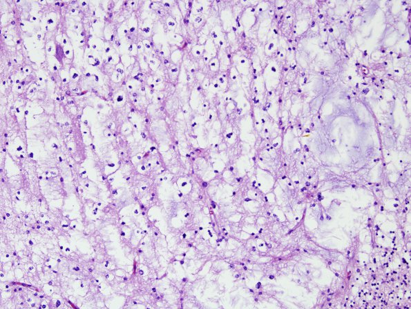
Washington University Experience | NEOPLASMS (NEURONAL) | DNET | 13A4 DNET, cerebellum (Case 13) H&E 3
Higher magnification of image #13A3 (H&E) ---- Additional data (not shown): Ki-67 shows a labeling index of <2% in areas corresponding to the neoplastic tissue. IDH1 is negative in neoplastic cells. Neurofilament highlights background axons. GFAP is positive in areas corresponding to neoplasm but does not stain the oligodendroglial cell-like component. P53 is negative. Synaptophysin highlights background neuropil. Neu-N highlights normal neurons of the cerebellar granular cell layer. ---- Comment: The findings are those of a low grade glioneuronal tumor, and are most consistent with DNT, WHO grade I.

