Table of Contents
Washington University Experience | NEOPLASMS (NEURONAL) | DNET | 15B DNET (Case 15) H&E
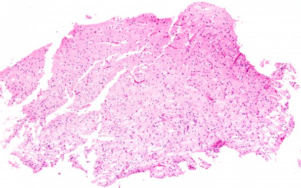
Minimal, minute specimen challenges the neuropathologist (H&E) --- Analysis (Scanned Images not available): Sections of the material from the left frontal lobe show a fragments of cortex with a suggestion of a mucin-rich tumor nodule. There is mild nuclear pleomorphism, with the majority of tumor nuclei being round and regular with bland chromatin and clear perinuclear halos. There are no Rosenthal fibers, or eosinophilic granular bodies. Mitotic figures are hard to find, and there is no evidence of endothelial hyperplasia or necrosis. The morphologic features are consistent with a low grade oligodendroglial tumor. The differential diagnosis would include both oligodendroglioma, WHO grade II and DNT, WHO grade I. FISH was performed revealing there were normal dosages (2 copies) of both chromosomes 1 and 19. No deletions were found. Since otherwise classic oligodendrogliomas demonstrate chromosome 1p and 19q deletions in roughly 85% of our cases, the lesion is most consistent with a DNT.

