Table of Contents
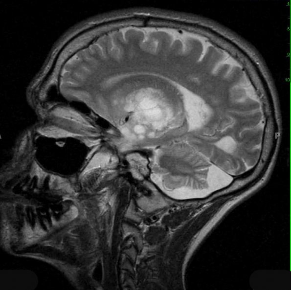
Washington University Experience | NEOPLASMS (NEURONAL) | DNET | 18A1 DNET (Case 18) T2 1 - Copy
Case 18 History ---- The patient is a 17 year old boy with a history of seizure disorder, who is status-post resection in 1996 of a left temporal lobe lesion diagnosed as DNT and has continuing seizures. He also has a history of osteochondroma of the left proximal medial tibia, excised in 05/2005. He now has a multicystic brain lesion involving the left temporal lobe and basal ganglia, which has increased in size since previous imaging was performed in 2001. ---- 18A1,2 The MRI cerebral lesion measures roughly 5.8 x 6.3 x 5.1 cm and shows distinct areas of nodular enhancement, one of which is new since 2001. Mild mass effect on the lateral ventricle and a 4 mm midline shift are also new since 2001. Operative procedure: Craniotomy.

