Table of Contents
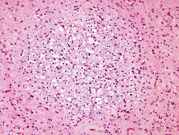
Washington University Experience | NEOPLASMS (NEURONAL) | DNET | 18B DNET (Case 18) H&E
Histological sections of the left temporal lobe lesion show fragments of a complex lesion. Focal areas with pilocytic features (including bipolar cells, eosinophilic granular bodies, Rosenthal fibers, and hyalinized microvascular proliferation with glomeruloid features in a myxoid background) are juxtaposed with areas exhibiting classic features of DNT including small round oligodendroglia-like cells, fine "chicken-wire" vasculature, pools of mucin often containing 'floating neurons,' and irregularly-shaped microcalcifications. Additionally, these ostensibly well-circumscribed DNT-like areas appear to transition into a mucin-free form as they involve the adjacent brain parenchyma, maintaining oligodendroglia-like cellular morphology and fine vascularization, but showing more abundant neuronal entrapment; this mucin-free form comprises the majority of specimen B, which show only focal DNT-like areas and lacks pilocytic-astrocytoma-like features. Mitotic figures are rare (<<1/2/40X) and necrosis is absent.

