Table of Contents
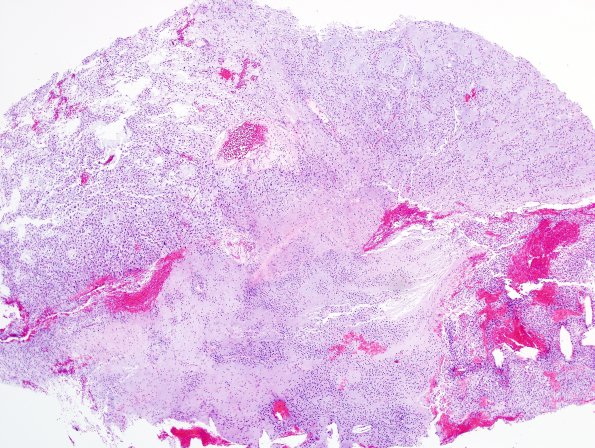
Washington University Experience | NEOPLASMS (NEURONAL) | DNET | 20A1 DNET (Case 20) H&E 21.jpg
Case 20 History ---- The patient is an 18 year old male with no clinical history provided other than a clinical diagnosis: Intractable complex partial seizure disorder. Operative procedure and findings: Second craniotomy for subdural grid removal and resection of (left parietal) seizure focus. 20A1-5 Microscopic examination of the sections shows neoplasm consistent with a DNT. The tumor is composed of round, hyperchromatic nuclei with indistinct cytoplasmic boundaries and clear cytoplasm and is moderately cellular with nodules of tumor cells separated by pools of mucinous-appearing material. Thin delicate capillaries with a chickenwire pattern are present. The tumor involves adjacent cortex and individual neurons are identified "floating" within the mucinous pools in the tumor. There is no evidence of increased cellularity, mitotic activity or necrosis within these sections.

