Table of Contents
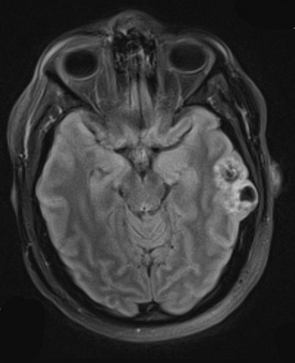
Washington University Experience | NEOPLASMS (NEURONAL) | DNET | 24A1 DNET (Case 24) TIRM 1 - Copy
Case 24 History ---- The patient is a 29-year-old man with a history of generalized tonic-clonic seizures starting in 2010. Radiologic imaging in 01/2011 identified a left temporal tumor which has since been monitored. ---- 24A1,2 MRI shows a ~ 3.7 cm complex cortically-based mass with calcifications, centered in the medial inferior left temporal gyrus. The lesion includes an anterior portion with a central hypointensity, and minimal enhancement, and a posterior portion that is cystic and has an enhancing mural nodule. The lesion remodels the inner table of the adjacent temporal calvaria. Radiological diagnosis: Most compatible with ganglioglioma or dysembryoplastic neuroepithelial tumor. Operative procedure: Left craniotomy and tumor resection with possible intraoperative MRI.

