Table of Contents
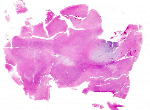
Washington University Experience | NEOPLASMS (NEURONAL) | DNET | 24B1 DNET (Case 24) H&E whole mount
24B1-3 Hematoxylin and eosin stained sections of the resection material show a complex, heterogeneous neoplasm with a nodular distribution within neocortex and white matter. The most defining 'specific glioneuronal element' appears as a ~0.5 cm nodule, centered at the gray-white junction, with abundant basophilic mucin, a web-like arrangement of delicate vasculature, a robust population of oligodendroglioma-like cells, and numerous entrapped cortical neurons. Nearby the nodule are several much smaller foci that contain similiar cytological elements, but lack basophilic mucin.

