Table of Contents
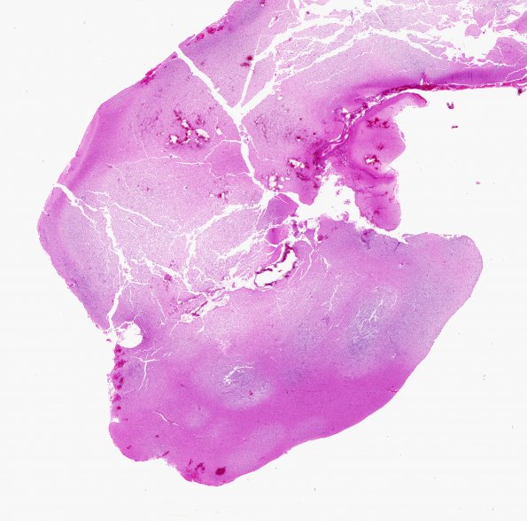
Washington University Experience | NEOPLASMS (NEURONAL) | DNET | 26A1 DNET (Case 26) H&E whole mount
Case 26 History ---- The patient was an 18 y/o male diagnosed with a DNT of the right parietal lobe in Dec 2004. Although clinically well, recent imaging in 2006 revealed a new enhancing nodule at the previous site of resection. A second surgery, with intraoperative MRI, appears to have resected the abnormality. ---- 26A1-3 Microscopic section from the 2004 and 2006 resections reveal a DNT, WHO grade I. The tumor is primarily cortically based and contains both a "patterned" nodular and non-nodular architecture. Numerous oligodendroglial-like cells are at times arranged in columns/trabeculae and are accompanied by neurons "floating" in mucin. Vasculature is capillary-like and delicate. There is no mitotic activity, microvascular proliferation (MVP) or necrosis. Microscopic sections from the 2006 biopsy reveal a predominance of more normal to mildly gliotic neocortex. However, rare small areas appear to contain abnormal tissue consistent with the prior surgical material and hence a DNT. It is impossible to determine if this material is truly "recurrent" or simply residual. ---- One small hypercellular fragment contains endothelial or microvascular proliferation (MVP), which may account for the imaging findings of an enhancing nodule. While MVP may be seen as part of DNT, this may also represent a reactive change adjacent to the tumor or to the prior surgical site. Deeper sections from the 'B' block did not reveal any additional tumor tissue.

