Table of Contents
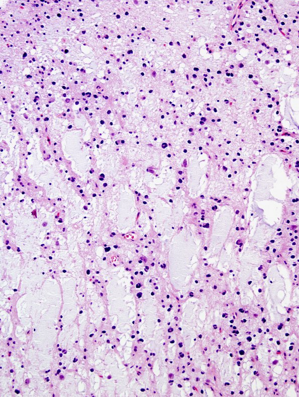
Washington University Experience | NEOPLASMS (NEURONAL) | DNET | 28A1 DNET (Case 28) H&E 4.jpg
28A1,2 Sections of the "right parietal lobe" biopsy material show a DNT which has a vaguely multinodular configuration. The small nodules are fairly well circumscribed and are composed of an admixture of glial cells and neurons freely floating in a myxoid/mucopolysaccharide-rich background. No mitotic figures are present. The nodules do not show infiltrative edges or satellitosis around the surrounding neurons.

