Table of Contents
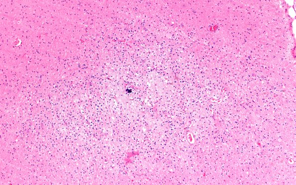
Washington University Experience | NEOPLASMS (NEURONAL) | DNET | 2B1 DNET (Case 2) A2 areaA H&E 10X
2B1-3 Orientation was optimal on this lesion perpendicular to the cortical surface. In many cases specimens are removed in fragments making orientation difficult and it behooves the neuropathologist to recognize elements of DNTs from fragments by histology, immunohistochemistry with support from imaging and clinical history. Tumor cells are set within large spaces made up mostly of background loosely textured/interwoven glial processes or mucin-rich extracellular material. The predominant cell population is oligodendrocyte-like tumor cells with uniform, relatively small oval to round nuclei containing evenly distributed chromatin and minimal mitotic activity. These cells are arranged in columns along axonal structures and occasional small blood vessels. Occasional 'floating neurons' are identified. Microcalcifications are present. This group of images are collected on the same microscopic lesion at 10X (2B1), 20X (2B2) and 40X (2B3). (H&E)

