Table of Contents
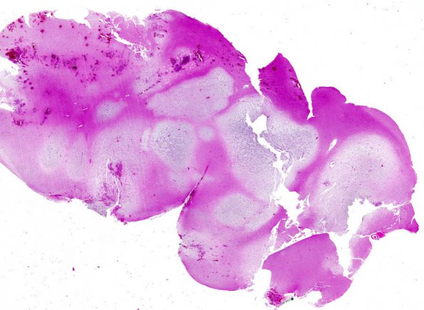
Washington University Experience | NEOPLASMS (NEURONAL) | DNET | 5A1 DNET (Case 5) H&E whole mount 1
Case 5 History The patient was a 21 year old woman with a history of intractable seizures for the last 6 years. MRI reveals a multi-cystic left temporal lesion with small punctate foci of enhancement. 5A1-4 Sections show a markedly expanded cortex with multiple mucin-rich microcystic nodules. These nodules have a patterned appearance with ribbons and linear arrays composed of oligodendroglial-like cells embedded in a gliofibrillary matrix. In between these nodules, the cortex is also hypercellular, containing predominantly oligodendroglial-like cells. There are scattered "floating neurons" within a small pool of mucin. A single mitotic figure is found. There is no evidence of either endothelial hyperplasia or necrosis.

