Table of Contents
Washington University Experience | NEOPLASMS (NEURONAL) | Desmoplastic Infantile Ganglioglioma (DIG) | 1A1 DIG (Case 1) - Copy
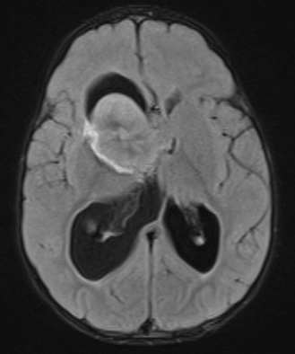
Case 1 History ---- The patient is a 2-year-old female with sickle cell disease who presented to the Hematology Service with limited use of her left arm. MRI revealed a 4.5 x 4.3 x 4.1 cm heterogeneously enhancing right hypothalamic mass with numerous foci of signal loss on susceptibility weighted images, a 2.7 cm homogenously enhancing component adjacent to the right lateral ventricle. Operative procedure: Right frontal craniotomy. ---- MRI Studies: 1A1 The mass is large, iso- to hyperintense to brain and appears to involve the base of the brain extending into the deep gray.

