Table of Contents
Washington University Experience | NEOPLASMS (NEURONAL) | Desmoplastic Infantile Ganglioglioma (DIG) | 2C4 DIG (Case 2) H&E 4
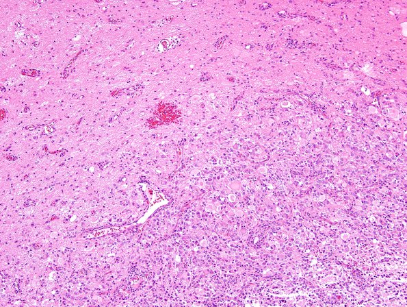
2C4-8 The tumor has a relatively sharp border with the underlying brain parenchyma, which shows numerous scattered atypical ganglion cells with marked cell size variation and multinucleation. Abundant axonal spheroids are also present in these areas. Another component shows smaller, more immature-appearing cells intermixed with gemistocytic astrocytes. Occasional mitotic figures are identified in this area. No necrosis or vascular proliferation is identified.

