Table of Contents
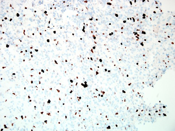
Washington University Experience | NEOPLASMS (NEURONAL) | Desmoplastic Infantile Ganglioglioma (DIG) | 3G DIG (Case 3) Ki67 2.jpg
IHC for proliferation marker Ki-67 (MIB-1 antibody) stains a regionally variable proportion of tumor cell nuclei, manually calculated in an area of estimated maximal density to be 17.5% which, in the presence of more than a few mitoses was worrisome, although increased mitoses may be seen in a patient of her age (4months). Nonetheless, the patient is alive and well 6 years later at her last MRI exam with no evidence of recurrence. ---- Ancillary tests (not shown): Neurofilament immunohistochemistry reveals entrapped axons at sparse-to moderate density in limited regions of tumor tissue, corresponding to areas with entrapped neurons; this finding is consistent with a relatively solid but also somewhat infiltrative growth pattern. IHC for CD34 highlights only endothelial cells; no dysmorphic neurons or highly branched cells are identified. IHC for BRAF V600E is negative. IHC for p53 highlights only a small subset of tissue nuclei (<5%).

