Table of Contents
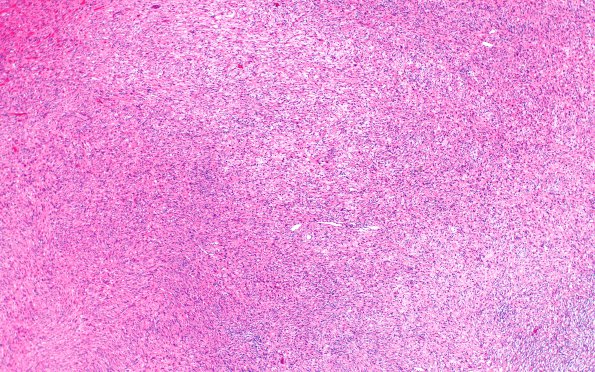
Washington University Experience | NEOPLASMS (NEURONAL) | Desmoplastic Infantile Ganglioglioma (DIG) | 4A1 DIG (Case 4) H&E 4X
Case 4 History ---- The patient is a 5 month old boy with normal development and who "consistently turned his head to the left," presented with one day of vomiting, diarrhea, and one episode of seizures. Imaging shows a large cystic lesion in the left parietal lobe with hemorrhage and a mural nodule. Operative procedure: Left parietal craniotomy and excision of cystic mass. ---- 4A1-4 Sections of the left parietal mass show a well-circumscribed, hypercellular neoplasm consisting predominantly of spindled epithelial cells with oval to elongated, hyperchromatic nuclei and moderate to abundant amount of eosinophilic cytoplasm. The neoplasm shows significant collagen and fibrosis admixed with tumor cells. The spindle cells are arranged in fascicles or in a vaguely storiform pattern with dilated vascular spaces. Mitoses are hard to find and there is no evidence of necrosis or vascular proliferation.

