Table of Contents
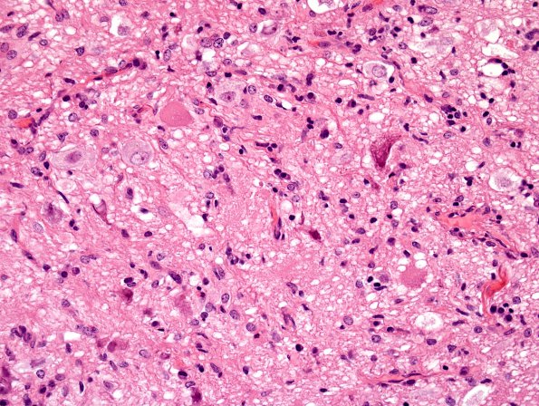
Washington University Experience | NEOPLASMS (NEURONAL) | Ganglioglioma | 11A1 Ganglioglioma (Case 11) H&E 4.jpg
Case 11 History ---- The patient is a 55-year-old woman with a seizure. Operative procedure: Craniotomy, amygdalohippocampectomy. ---- 11A1,2 H&E stained sections of the resection specimen show a low-grade neoplasm. Both neoplastic neuronal (i.e., ganglionic) and glial forms are present. In some areas, abnormal neurons are found in clusters and other unusual arrangements. Numerous clear vacuoles are seen in the neuropil and in some neurons. There are focal perivascular lymphocytic infiltrates. Mitotic activity is minimal or non-existent. Microvascular proliferation and necrosis are also absent.

