Table of Contents
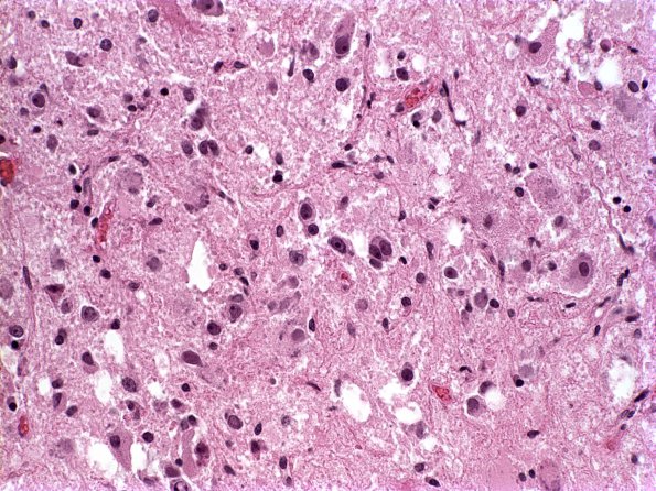
Washington University Experience | NEOPLASMS (NEURONAL) | Ganglioglioma | 13A1 Ganglioglioma (Case 13) H&E 1
Case 13 History ---- The patient is a 42 year-old man with a history of parietal tumor, s/p resection at the University of Missouri-Columbia Hospital and Clinics in July of 1999. ---- 13A1,2 H&E slides show a low grade glioneuronal tumor. Interspersed among the glial tissue are single or clustered neurons with vesicular nuclei, prominent nucleoli and abundant cytoplasm with Nissl substance. Some of these neurons have dysmorphic features characterized by binucleation or eccentricity of irregular nuclei. The glial neoplastic component is composed of cells with oval to spindled hyperchromatic nuclei and indistinct or stellate cytoplasmic processes, consistent with astrocytes which are arranged haphazardly or in intersecting fascicles. Rosenthal fibers and eosinophilic granular bodies are identified. Mitotic figures are hard to find. There is no necrosis or endothelial hyperplasia.

