Table of Contents
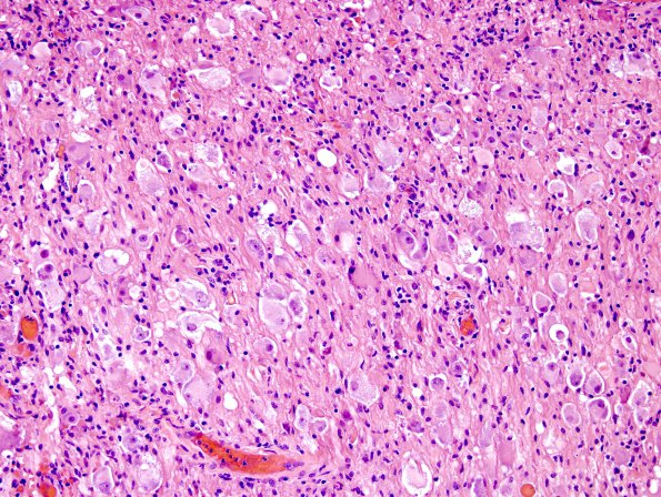
Washington University Experience | NEOPLASMS (NEURONAL) | Ganglioglioma | 14A1 Ganglioglioma (Case 14) H&E 5
Case 14 History ---- The patient is a six year old boy with a brainstem mass. Operative procedure: Craniotomy for excision of posterior fossa tumor. ---- 14A1-3 Sections of the "posterior fossa tumor," show a moderately cellular neoplasm composed of large tumor cells with abundant eosinophilic cytoplasm, rounded nuclei and prominent nucleoli in a fibrillary background. Many cells also contain Nissl substance, all features consistent with ganglion cell differentiation. Numerous swollen processes with glassy cytoplasm are consistent with axonal spheroids. Smaller cells with elongate, mildly hyperchromatic nuclei are also seen, consistent with an astrocytic component. Mitotic figures are hard to find. There is perivascular inflammation and hemosiderin laden macrophages are present.

