Table of Contents
Washington University Experience | NEOPLASMS (NEURONAL) | Ganglioglioma | 16A2 Ganglioglioma (Case 16) H&E 4.jpg
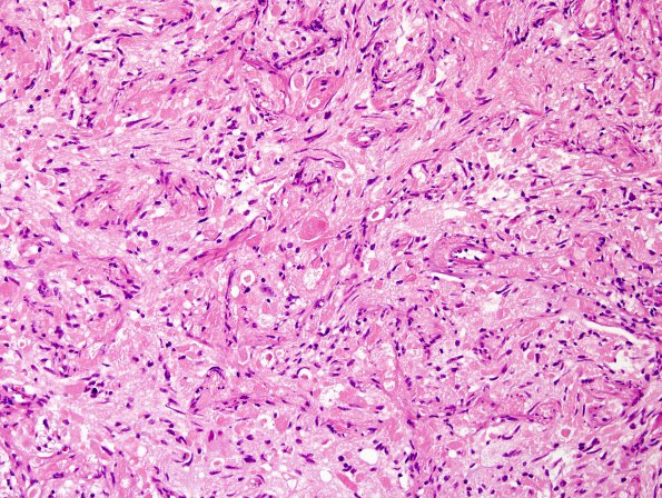
16A2-4 This is a moderately cellular low grade glioneuronal neoplasm with abundant amounts of eosinophilic granular bodies found throughout the tumor. The tumor has a predominantly solid growth pattern. Focally, there are islands of collagen fibrosis within the tumor and a minute focus of bland necrosis. The tumor has two major cellular components; neuronal and glial. There are scattered dysmorphic appearing neurons with enlarged and multipolar cell bodies and coarse Nissl substance. The glial elements show mild to moderate pleomorphism and have a heterogenous appearance. There are some areas where the glial cells have a spindle cell appearance and are arranged in fascicles. The glial nuclei are elongated to spindled, irregular, and have hyperchromatic to vesicular chromatin patterns. Mitotic figures are not readily identified. Portions of the "cyst wall" show reactive changes including piloid gliosis with Rosenthal fiber deposition.

