Table of Contents
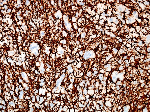
Washington University Experience | NEOPLASMS (NEURONAL) | Ganglioglioma | 19F Ganglioglioma (Case 19) GFAP 3.jpg
GFAP immunohistochemistry for glial fibrillary acidic protein is strongly positive within most of the smaller tumor cells and a feltwork of small processes, and negative within the ganglion cells. ---- Ancillary data (not shown): IHC for NeuN highlights sparse, relatively mature-appearing neurons (consistent with entrapped non-neoplastic neurons) with strong intensity, and only a questionably neoplastic subset of the ganglion cells with modest intensity. Histochemical periodic acid Schiff stain highlights eosinophilic granular bodies and some of the ganglion cells. IHC for CD34 highlights only endothelial cells; no highly branched parenchymal forms are detected. BRAF V600E is negative. IHC for proliferation marker Ki67 (MIB-1 antibody) stains a regionally variable proportion of tumor cell nuclei, ranging up to 11.7%.

