Table of Contents
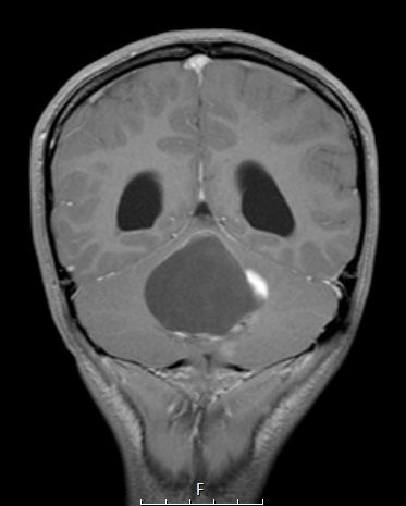
Washington University Experience | NEOPLASMS (NEURONAL) | Ganglioglioma | 21A Ganglioglioma (Case 21) T1W 1 - Copy
Case 21 History ---- The patient is a 15-year-old girl with a four month history of headache and gait instability. MRI showed a 5.1 cm intra-axial cystic mass in the midline cerebellum with peripheral nodular enhancement. She underwent resection; examination of the resected material yielded the diagnosis of 'pilocytic astrocytoma, WHO grade I. Postoperative MRI showed areas of residual enhancement suspicious for residual tumor. Operative procedure: Posterior fossa craniotomy for resection of residual tumor. ---- 21A T1-weighted scan prior to her first biopsy with contrast shows a cyst with a mural nodule in the cerebellum. This MRI was thought more likely to represent a pilocytic astrocytoma than a ganglioglioma in terms of numbers alone. In this case the biopsy the tumor was thought to be a pilocytic astrocytoma; however, resection showed a ganglion cell component not present in the first biopsy.

