Table of Contents
Washington University Experience | NEOPLASMS (NEURONAL) | Ganglioglioma | 3B1 Ganglioglioma (Case 3) H&E 2
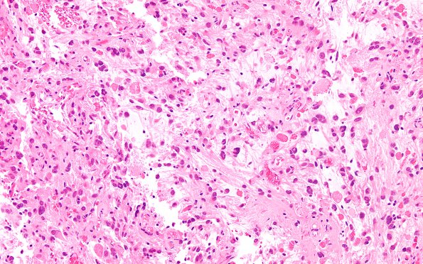
3B1-3 Routine H&E stained sections show a cellular neoplasm with both dense fibrillary and loose microcystic areas with both glial and gangliocytic components. The tumor cells in the glial component have oval nuclei with dedicated hair-like “piloid" processes, while other tumor cells have round nuclei with inconspicuous nucleoli. There are occasional multinucleated tumor cells with nuclei arranged at the periphery of the cells. Large dysplastic ganglion cells, some bi- or multi-nucleate, are present. There is abundant dedicated branching vasculature with no compelling endothelial proliferation. The vessel walls are focally hyalinized. There are frequent Rosenthal fibers and eosinophilic granular bodies. There is no significant mitotic activity or necrosis.

