Table of Contents
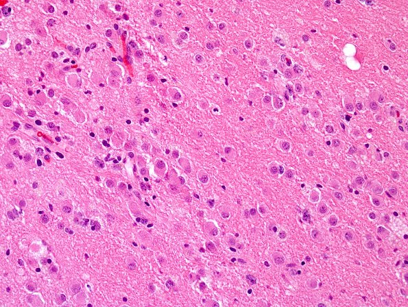
Washington University Experience | NEOPLASMS (NEURONAL) | Ganglioglioma | 5A1 Ganglioglioma-gangliocytoma (Case 5) H&E 1.jpg
Case 5 History ---- The patient is a 63-year-old woman with remote history of viral encephalitis who recently developed blurred vision, headache, and bilateral upper extremity heaviness. MRI from July 2016 showed a T2 hyperintensity within the white matter of the right temporal lobe associated with thinning of the overlying cortex and sulcal enlargement. Operative procedure: Right craniotomy and brain biopsy. ---- 5A1-3 The temporal lobe mass showed a low-grade primary central nervous system neoplasm made up predominantly of small, monomorphic mature-appearing neurons with moderate amounts of cytoplasm, eccentrically located, mildly atypical nuclei with occasional contour irregularity and binucleation. Neoplastic neurons are overly abundant but irregularly distributed within the background parenchyma, frequently appearing within abnormal clusters.

