Table of Contents
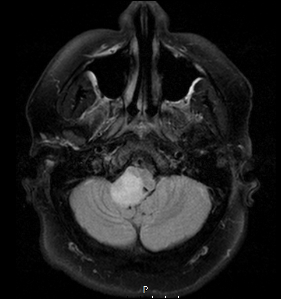
Washington University Experience | NEOPLASMS (NEURONAL) | Ganglioglioma | 6A1 Ganglioglioma (Case 6) FLAIR 1 - Copy
Case 6 History ---- The patient is a 19-year-old man who had been recently diagnosed with severe sleep apnea and also experienced some diplopia, occasional blurry vision, and unsteady gait. MRI showed a 5.4 x 2.8 x 2.6 cm expansile, T2 hyperintense, enhancing lesion centered in the right posterolateral medulla, with extension superiorly into the right middle cerebellar peduncle, inferiorly into the upper cervical cord, and posteriorly (where it appears exophytic) into the right foramen of Luschka. The lesion also produces mass effect on the inferior aspect of the right cerebellar tonsil. Radiological differential: Primary glial tumor such as pilocytic astrocytoma and low grade glioneuronal tumor; lymphoma; less likely, tumefactive demyelination. Spinal MRI showed no evidence of 'drop' metastasis. Operative procedure: Midline posterior craniotomy for biopsy and partial resection. ---- MRI Studies: 6A1 The precarious location of this tumor is shown in this FLAIR scan.

