Table of Contents
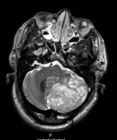
Washington University Experience | NEOPLASMS (NEURONAL) | Lhermitte-Duclos Disease | 1A Lhermitte-Duclos Dz (Case 1) T2W - Copy
Case 1 History ---- This was a 47-year-old man with a history of left posterior fossa craniotomy for tumor resection in 1998 (final diagnosis unknown). He recently presented following a fall and altered mental status. A head CT scan showed fourth ventricle effacement, posterior fossa edema, and evidence of tonsillar herniation. Brain MRI showed a large expansive T2 hyperintense mass centered in the left cerebellar hemisphere with a striated appearance, and significant associated mass effect. Operative procedure: Posterior fossa craniotomy for tumor resection. ---- This T-2 weighted MRI scan shows a “tiger stripe pattern” of the cerebellar lesion.

