Table of Contents
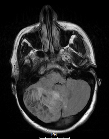
Washington University Experience | NEOPLASMS (NEURONAL) | Lhermitte-Duclos Disease | 2A1 Lhermitte-Duclos (Case 2) FLAIR - Copy
Case 2 History ---- The patient was a 32-year-old woman with a right cerebellar mass, with imaging suspicious for Lhermitte-Duclos syndrome. The patient has a vague history of headaches, intermittent blurry vision, and tingling of the arms. At times, the patient has had trouble with fine motor tasks such as writing. MR imaging performed at BJH which showed a lesion centered at the right cerebellar hemisphere with high T2 signal measuring up to 6.6 cm, causing mass effect on the fourth ventricle and cervicomedullary junction. Operative procedure: IMRI with right craniotomy for tumor. ---- 2A1-5 MRI examination: 2A1 This FLAIR scan shows patchy hyperintensity adjacent to a hypointensive mass lesion.

