Table of Contents
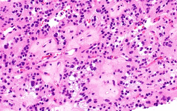
Washington University Experience | NEOPLASMS (NEURONAL) | Papillary Glioneuronal Tumor (PGNT) | 1B3 Papillary Glioneuronal Tumor (PGNT) (Case 1) H&E 3
1B2-5 Most tumor cells surround pale hyalinized vessels, exhibiting a pseudo-rosette pattern. They have round to oval nuclei with various sizes including some enlarged hyperchromatic nuclei, indistinct micro-nucleoli, fibrillary eosinophilic cytoplasm with some microvacuolation. Some areas exhibit vague microcyst patterns. Mitoses are not seen. There is no definitive evidence of atypia, microvascular endothelial proliferation or tumor necrosis. (H&E)

